
In the realm of medical diagnostics, few tests offer the insight and clarity that echocardiography, or echo, brings to the examination of the heart. It's a test that transcends pain, utilizing sound waves to create a visual symphony of your heart's inner workings. But what exactly is echocardiography, and when might you need it?
Echocardiography Unveiled
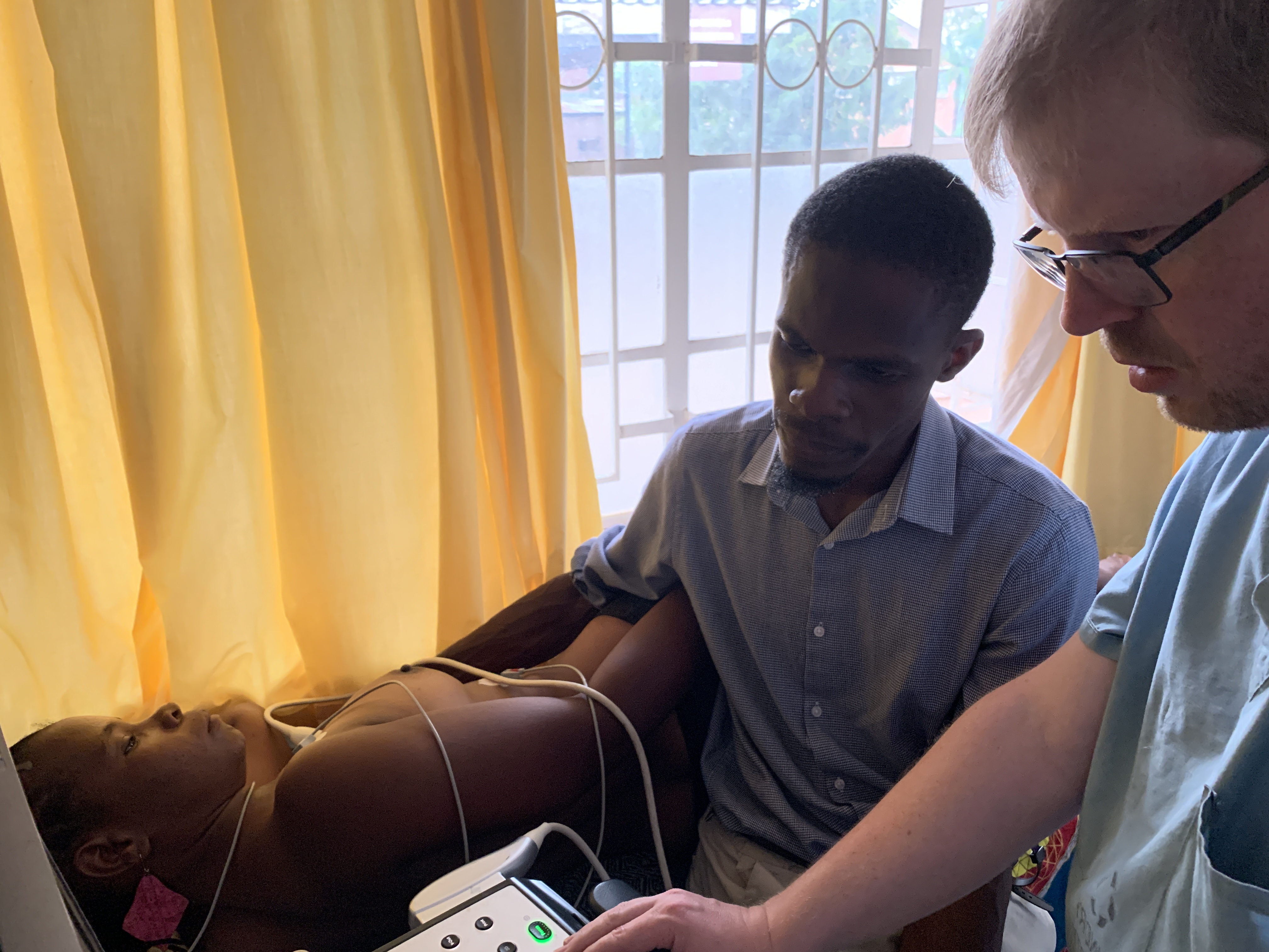
Echocardiography, lovingly known as echo, is a painless and non-invasive test that harnesses the power of sound waves to produce moving images of your heart. These images reveal not only the size and shape of your heart but also the precise functionality of its chambers and valves.

Diving Deeper with Doppler
Echo goes beyond the surface, delving into the intricacies of blood flow within your heart's chambers and valves, thanks to a special type of echo known as Doppler ultrasound. It unveils how efficiently your heart pumps blood and can detect potential issues like blood clots within the heart, pericardial fluid accumulation, or aortic anomalies.
Echo's Multifaceted Role
Who benefits from echocardiography? Essentially, anyone with signs or symptoms of heart problems may find solace in the insights echo provides. For example, shortness of breath and leg swelling may hint at heart failure, a condition where the heart struggles to pump enough oxygen-rich blood to meet the body's needs. Echo steps in to reveal the heart's pumping efficiency.

Unveiling Heart Mysteries
But echo does more than just uncover heart failure; it's a diagnostic detective, deciphering the mysteries of abnormal heart sounds like murmurs. These murmurs can be harmless or, in some cases, signals of underlying heart problems.
A Glimpse into the Heart's Inner Workings
Echo offers an intricate look into various aspects of your heart's health:
Varieties of Echo
Echocardiography isn't a one-size-fits-all test; it comes in various forms:
The Echo Experience
Undergoing an echocardiography is straightforward. There are no special preparations required for most echo types. You can eat, drink, and take medications as usual. Some exceptions apply, such as fasting before a TEE.
During the test, electrodes are placed on your chest, an EKG is performed, and a gel is applied to enhance ultrasound wave transmission. A transducer is then moved across your chest to capture images, often taking less than an hour.
For TEE, you might receive a mild sedative, and the transducer is inserted into your esophagus. Recovery from TEE may involve monitoring for a few hours.

Echo's Role in Your Heart's Story
Echocardiography doesn't just provide images; it offers answers and insights. It's a diagnostic dance with the heart's rhythm, revealing tales of health and potential concerns. While no medical test is without its considerations, echo remains a vital tool in deciphering the heart's mysteries, offering clarity, and guiding the way toward a healthier tomorrow.
Sources
